
Advanced Upper Cervical Spine Stabilization by Dr. Alok Gadkari
Understanding the C1–C2 Joint (Atlas–Axis Joint)
The C1 vertebra (Atlas) supports the skull, while the C2 vertebra (Axis) contains a bony projection called the odontoid (dens), allowing head rotation.
This joint is responsible for:
Head rotation (looking left and right)
Stability of the skull on the spine
Protection of the spinal cord and brainstem
Because of its mobility and anatomical complexity, instability at C1–C2 can be dangerous if not treated properly.
What is C1–C2 Instability?
C1–C2 instability occurs when there is excessive movement between the Atlas and Axis vertebrae. This abnormal movement can compress the spinal cord or brainstem, leading to neurological complications.
Common Causes
Trauma or fracture (odontoid fracture)
Road traffic accidents
Rheumatoid arthritis
Congenital abnormalities
Ligament injury
Degenerative conditions
Tumors or infections
Down syndrome (in children)
Early diagnosis and proper stabilization are crucial to prevent permanent neurological damage.
Symptoms of C1–C2 Instability
Symptoms vary depending on severity. Some patients may experience only neck pain, while others develop neurological deficits.
Common symptoms include:
Severe upper neck pain
Restricted neck movement
Headache at the base of skull
Tingling or numbness in arms
Weakness in limbs
Difficulty walking
Loss of coordination
Balance problems
In severe cases, breathing difficulty
If left untreated, instability can cause spinal cord compression, which may result in paralysis.
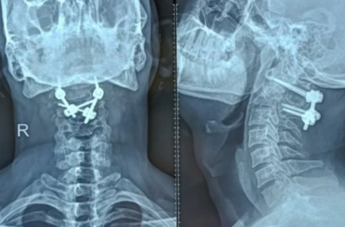
What is C1–C2 Fusion Surgery?
C1–C2 Fusion is a surgical procedure that permanently joins the first and second cervical vertebrae to eliminate abnormal motion and stabilize the spine.
The procedure involves:
Placement of screws in C1 and C2 vertebrae
Connecting rods to secure stability
Bone graft placement to promote fusion
Over time, the bones grow together, forming a solid union that prevents dangerous movement.
Goals of C1–C2 Fusion Surgery
Stabilize the upper cervical spine
Relieve spinal cord compression
Reduce neck pain
Prevent neurological deterioration
Protect brainstem function
Restore patient mobility and confidence
Why Choose Dr. Alok Gadkari for C1–C2 Fusion?
C1–C2 fusion is a technically demanding surgery requiring precision and experience. Dr. Alok Gadkari specializes in complex spine procedures with a focus on:
Accurate diagnosis
Advanced imaging guidance
Modern instrumentation techniques
Minimizing surgical risks
Patient-centered care
Structured rehabilitation protocols
His expertise in cervical spine surgery ensures optimal safety and recovery outcomes.
Diagnostic Evaluation Before Surgery
Proper diagnosis is critical for planning surgery.
Dr. Alok Gadkari may recommend:
X-rays (dynamic flexion-extension views)
MRI scan (to assess spinal cord compression)
CT scan (for bone anatomy)
Neurological examination
Blood investigations
Imaging helps determine instability severity and the best surgical technique.
Surgical Techniques Used in C1–C2 Fusion
Modern techniques have significantly improved safety and outcomes.
1. Posterior C1–C2 Screw Fixation
This is the most common method.
Screws are placed in C1 lateral mass
Screws are placed in C2 pedicle or pars
Rods connect the screws
Bone graft promotes fusion
2. Transarticular Screw Fixation
Screws are placed across C1–C2 joint
Provides strong fixation
Requires precise anatomical alignment
3. Occipito-Cervical Fusion (if needed)
If instability extends upward to skull base, fusion may include occiput.
Dr. Alok Gadkari selects the most appropriate technique based on patient anatomy and pathology.
How is the Surgery Performed?
General anesthesia is administered
Patient positioned carefully to protect spinal cord
Small posterior incision made
Precision-guided screw placement
Rod fixation applied
Bone graft inserted
Wound closed carefully
Surgery typically takes 2–4 hours depending on complexity.
Benefits of C1–C2 Fusion Surgery
Prevents spinal cord injury
Reduces severe neck pain
Improves neurological symptoms
Stabilizes unstable fractures
Enhances quality of life
Allows safe return to daily activities
Though neck rotation reduces slightly, most patients adapt well and maintain functional mobility.
Recovery After C1–C2 Fusion
Immediate Post-Operative Phase
Hospital stay: 3–5 days
Pain management
Neck support brace (if required)
Early mobilization
First 6 Weeks
Restricted heavy lifting
Gradual increase in walking
Follow-up imaging
Wound care monitoring
3–6 Months
Bone fusion develops
Physiotherapy improves strength
Return to work (depending on occupation)
Full fusion may take 6–12 months.